
Internal Medicine
A true specialization of general medicine, dealing with all pathologies.
Internal medicine is the specialty of difficult diagnostic approaches and the management of animals suffering from polypathologies or general diseases.
Tumors of the spleen

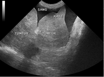
Spleen tumors represent 7% of tumors encountered in dogs. They are much rarer in cats.
The most common tumor of the spleen is hemangiosarcoma (called splenic hemangiosarcoma). Certain breeds are predisposed to it: the German Shepherd, the Golden Retriever, the Labrador, the Boxer, the Poodle… but can be found on all breeds with a predominance in medium to large breeds.
When it comes to malignant tumors, which represents about one in two cases, their prognosis is often poor. Indeed, the strong vascularization of the spleen means that these tumors tend to metastasize rapidly.
A mass on the spleen can sometimes be felt by your veterinarian during abdominal palpation. It is also possible that such a mass is visualized on X-ray, ultrasound or CT scan. However, most of the time, for lack of preventive examinations or evocative symptoms, the masses on the spleen are not detected until late, when this organ ruptures. An abdominal hemorrhage is then present. The animal is usually very downcast, in shock, has pale mucous membranes, may have breathing difficulties and a distended abdomen. In this case, surgery will be performed urgently, with the primary objective of stopping the bleeding. If necessary, additional examinations and an extension assessment (blood test, abdominal and/or cardiac ultrasound, chest x-rays, etc.) may be performed later.
In the presence of a splenic mass, surgery is required. It is only after histological analysis that the nature of the mass can be determined and therefore the possible diagnosis of splenic hemangiosarcoma established. If this has not been done prior to surgery, the extension assessment mentioned above makes it possible to know whether or not there are already metastases and therefore to refine the prognosis (which is of course clouded by the proven presence of metastases). The decision of a chemotherapy protocol can then be taken.
Due to the crude or not very suggestive clinical symptoms, splenic hemangiosarcomas remind us of the importance of additional examinations as a preventive measure in our dogs from 6 years of age. Ideally, hematology and ultrasound of the spleen should be performed every year.

